Our Mission

The objective of the GlyCORE Imaging Core is to promote and enhance the growth of glycoscience projects at the University of Mississippi and throughout the mid-south region. The Core brings together new and existing advanced microscopes into a University-wide central platform, offering a wide range of advanced imaging techniques, including laser scanning confocal microscopy and related beam parking experimental approaches, bright field and phase contrast microscopy, and computer-aided image analysis. The investigators will have access to and training in the use of advanced microscopy instrumentation, and assistance in the analysis of the resulting images. These images will provide new data and an understanding of the roles of diverse carbohydrates in living systems.

As many advanced microscopic techniques are new to the University of Mississippi campus, the Imaging Core will develop a training program to inform and train new users on these new techniques. These techniques include, but are not limited to, the live cell techniques of FRET, FLIP, and FRAP. Moreover, the GlyCORE Imaging Core will hold training sessions on how to properly get the most out of image analysis with instruction on the Leica Application Suite software, Image J/Fiji, the FARSIGHT toolkit for multi-dimensional microscopy, and Adobe Photoshop.

Imaging is essential for modern biological questions, and the GlyCORE Imaging Research Core will ensure glycoscience researchers at the University of Mississippi have access to these tools. These instruments and training sessions will also provide GlyCORE investigators with a partner in advanced imaging techniques.


Our Microscopes

Reserve a microscope here!

This microscope is equipped with a motorized Super Z stage, motorized water cool turret, and 10X, 20X, 40X, and 63X high NA objectives, capable of DIC and IR imaging.  It is equipped with three laser systems for excitation: i) a white light laser (WWL2), ii) a 405 nm DMOD laser, and iii) a 355 nm violet HeNe laser. This confocal also has the AOBS for tunable excitation for up to eight lines simultaneously, all with very step transitions (<5nm).  This is an extremely bright and spectrally flexible beam splitter that will allow for tremendous synergism with the white light laser.   Also equipped with three HyD detectors and a resonant scanner, this confocal microscope is both extremely fast and sensitive.  This confocal also has a powerful workstation with several software modules, including: i) LAS X system for control of microscope hardware, ii) Live Data Mode, iii) 3D visualization tool, iv) 3D Analysis tools, v) Wizards for FRAP, FLIP, and FRET, and vi) Hyvolution for super-resolution down to 140nm.

The Leica SP8 X imaging system coupled to the DMI8 epi-fluorescence microscope equipped with various components as listed below-

  • Objective lenses: 10X/0.40, 20X/0.70, 40X/1.25 (Oil, Ph), 63X/1.20 (Water), 63X/1.40-0.60 (Oil).
  • UV laser 405 nm for imaging UV fluorophores and photoactivation.
  • Laser 442 for imaging CFP, AF 430, etc.
  • The white light laser gives any wavelength from 470 nm to 670 nm in 1 nm increments.
  • Spectral Imaging
  • Six PMT detectors (5 fluorescence channels (3 conventional PMT and 2 hybrid PMTs) + 1 transmission channel).
  • resonant scanner (up to 8000 Hz).
  • Temperature-controlled incubator.

JEOL JSM-7200FLV Field-Emission Scanning Electron Microscope (FESEM)

This advanced microscope with high magnification and ultrahigh spatial resolution capabilities enables high-quality imaging and analysis of micro and nanostructures. It is equipped with multiple detectors, including an Energy Dispersive X-Ray Spectrometer (EDS), Scanning Transmission Electron Microscopy (STEM), Cathodoluminescence (CL), Back-Scattered Electron (BSE), and Secondary Electron (SE) detectors, enabling advanced analysis of various sample types.

SEM Core instrumentation

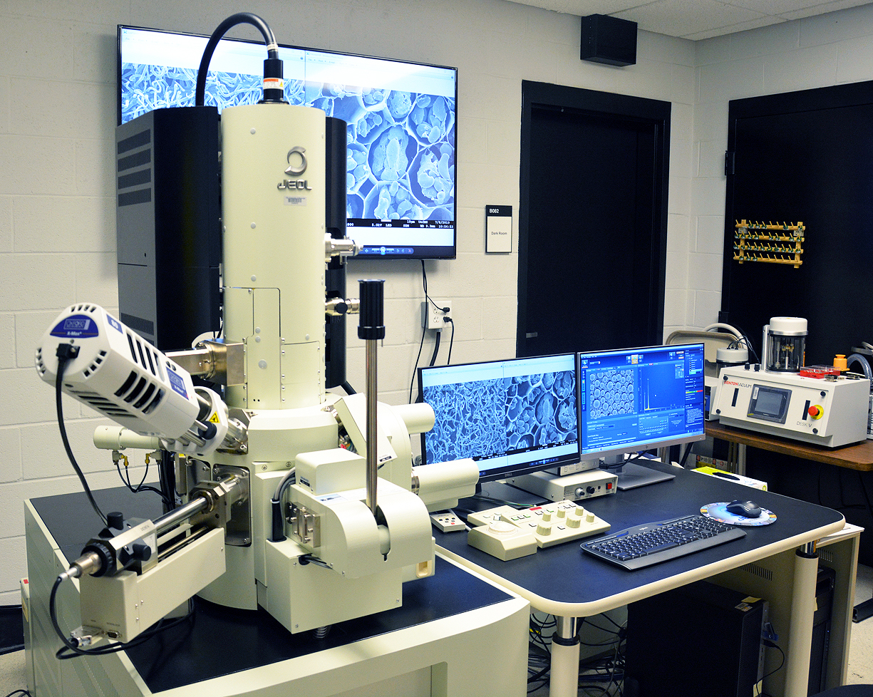
JEOL JSM-7200FLV FESEM at the Microscopy and Imaging Center

Specifications of the FESEM:

          Accelerating voltage            0.01 kV to 30 kV

          Probe current                        1 pA to 300 nA

          Magnification                        Up to x1,000,000

          Resolution                              Up to 1.2 nm

Detectors

  1. Secondary Electron (SE) detector
    • Everhart-Thornley type in-chamber Lower electron detector (LED), and
    • Through-the-lens (TTL)/Upper electron detector (UED)
  2. Back-Scattered Electron (BSE) detector
    • Retractable BE detector (RBED) with COMPO and SHADOW modes
  3. Energy Dispersive X-Ray Spectroscopy (EDS) detector
    • Oxford Instruments X-MaxN 80 EDS with AztecEnergy 5.0 software
  4. Scanning Transmission Electron Microscopy (STEM) detector
    • Deben Retractable motorised Annular STEM, HAADF/MAADF/LAADF/BF, with 12 position STEM converter holder for 3mm TEM grids
  5. Cathodoluminescence (CL) detector
    • Deben Centaurus monochromatic CL detector

Other Features

  • Low vacuum (LV) capability
  • Gentle Beam (GB) mode
  • Charge-free (CF) scan
  • Schottky-type thermal field emission gun
  • 5-axis, motorized stage
  • Trackball, touchpad, mouse and keyboard operation
  • Stage navigation system
  • Chamber scope
  • 65″ LCD display
  • Windows 10 Professional Operating System
  • Film thickness monitor
  • Data backup
  • Uninterruptible power supply

Denton Desk V TSC Sputter Coater

Denton Vacuum Desk V_TSC sputter coater

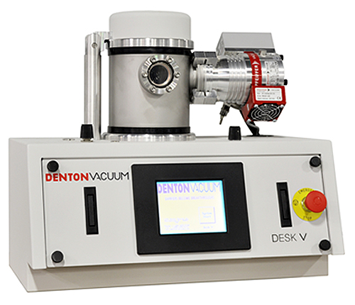
The Desk V sputter coater is used to prepare samples for SEM analysis. This fully automated sputter coater is designed for high-resolution electron microscopy. It is equipped with metal sputtering (Platinum) and carbon evaporation capabilities, oil-free turbo molecular pump, film thickness monitor and tilting/rotating specimen table for uniform coating. The unit is supplied with argon gas.

Leica EM CPD300 Critical Point Dryer

Leica EM CPD300 critical point dryer

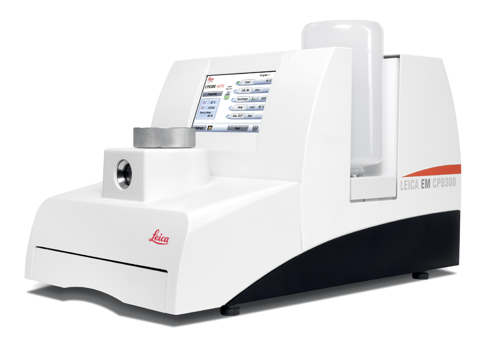
A critical point dryer is essential for preparing delicate biological specimens for SEM imaging. The Leica EM CPD300 is an advanced, fully automated unit capable of drying delicate materials for high-quality imaging. This unit uses liquid CO2(critical point 31.1 °C at 1072 psi) as a transitional medium.

Rates

User Fee

1

Services

UM Rate

External Academic Rate

Non-Academic Rate

Sample preparation, SEM imaging and EDS analysis

$75/hr

$110/hr

$150/hr (Minimum 2 hrs)

Hands-on training on SEM operation and sample preparation (min. 4 hrs)

$75/hr

$110/hr

On request

Demonstration for student groups (4-5 students per group)

$75/hr

$110/hr

On request


General Policy

Before you use any instrument in the facility for the first time, you need to have a consulting tour and discussion with the core staff.
New users must request/submit an account form for approval, and approved users will have a user account.

As one of the core facilities’ aims is to encourage learning instruments and self-use, training is highly recommended. If the Core Manager assists in the experimental design, data acquisition, and scientific interpretation, they should be recognized as a co-author in the research outcomes.

Users will be charged $75/per hour for internal users (UoM) and $110/per hour for external academic users.

The rate for non-academic users is $150/per hour with a minimum of two hours. This includes assistance from lab personnel setting up and operating the SEM.

The costs of essential consumables (aluminum stubs, carbon tapes, etc.) are included in the fee. The users are responsible for any other special materials required for preparing their samples.

Booking Policy

Reservation of the instruments before use is required. The billing is based on the length of occupation time, which is figured out from the reservation and the actual log-in and out time. Please record the log-in and log-out times in the reservation system. It also helps to track the usage of instrument components.

During reserved time, the lab is unavailable for others. The reserved person can use the lab to set up the room and protocol, sample preparation, image analysis, and clean up. Users are encouraged to finalize their work within the time reserved and leave the lab promptly so that the next user can use the lab immediately.

Please let us know in advance if you are unable to use the SEM at the reserved time so others can use it.

Invoicing and Payment Policy

Invoices for services provided in the previous month will be emailed to the investigator’s fiscal contact during the second week of the month.
Payments are due within 30 days of the invoice date. If payment is not received within three billing cycles, the user cannot make new reservations until all the pending invoices are paid in full.

We strive to determine the most efficient approach to achieving each user’s research goals and will make our best effort to generate high-quality, reliable results. However, we cannot guarantee that we can produce particular results in any research project. Payment is expected for services rendered, not contingent on a positive experimental outcome.

  • Motorized 8-100 zoom
  • Seamless integration into the modular system of SteREO microscopes
  • Motorized components for reliable results
  • Illumination and contrast methods based on cold light or LED
  • Excellent depth perception, even at high magnification levels
  • Ergonomic and easy operation and control with the optional touch panel SYCOP

Specs

Processor: (Single Gold 5xxx) Intel Xeon Gold 5218R 2.1GHz,(4.0GHz Turbo, 20C, 10.4GT/s 2UPI, 27.5MB Cache, HT (125W) DDR4-2666)

Graphics Card: (Nvidia single) Nvidia Quadra RTX 400, 8GB, 3DP, Virtual Link (XX20T)

Memory (standard memory) 48GB 6x8GB DDR4 2666MHz RDIMM ECC memory)

Operating System (Boot) Drive: Intel NVMe PCIe SSD (Front PCIe FlexBay)

Storage Drive Controllers: Intel Integrated controller (RST-e) with 1-2 Front FlexBay NVMe PCIe Drives

Hard Drive: (Class 40 PCIE SSD) M.2 2TB PCIe NVMe Class 40 Solid State Drive

2nd storage Drive: (Class 40 PCIE SSD) M.2 2TB PCIe NVMe Class 40 Solid State Drive

Software

Fiji

Photoshop

Zen Lite

LAS X

One Imaging channel (Brightfield)

Two Fluorescence Channels :(Ch1=>650 nm, Ch2=560-590 nm) One excitation wavelength (532 nm)

GlyCORE Imaging Core Inventory

Please be advised that these reagents are only for GLyCORE users and only for temporary tests; everyone should use their own reagents for regular experiments.

Secondary Antibodies
FluorophoreRRIDStore locationAntibodiesEx/EmAmount
AF488AB_26332754 degree Fridge (3rd Floor)Goat anti-Mouse488/5182x1mg
AF568AB_105635664 degree Fridge (3rd Floor)Goat anti-Rabbit575/6032x1mg
AF647AB_27628454 degree Fridge (3rd Floor)Goat anti-Chicken649/6701x1mg
AF405N/A4 degree Fridge (3rd Floor)Goat anti-Rat405/4201x1mg
Nuclear Staining
ProductRRIDStore LocationEx/EmApplication
NucRed™ Live 647N/A4 degree Fridge (3rd Floor)638/686Permeable membrane/Live
Hoechst 34580N/A4 degree Fridge (3rd Floor)392/440Permeable membrane/Live
DAPIAB_23074454 degree Fridge (3rd Floor)358/461Non- Permeable/Fixed
Actin Staining
ApplicationManualStore LocationProductEx/Em
F-actin stainingLink Dark Drawer (Imaging Core Office)CellMask Green Actin tracking stain503/512
Mitochondria Staining
ProductRRIDStore LocationEx/EmManual
MitoTracker™ Red CMXRosN/A-30 degree Fridge (3rd Floor)579/599Link
Lectin Staining Kit
ProductsRRIDStore LocationComponents
Lectin Kit IAB_23362524 degree Fridge (3rd Floor)Concanavalin A
Glycine max (soybean) agglutinin
Triticum vulgaris (wheat germ) agglutinin
Dolichos biflorus agglutinin
Ulex europaeus agglutinin I
Ricinus communis agglutinin
Arachis hypogaea (peanut) agglutinin
Lectin Kit IIAB_23362534 degree Fridge (3rd Floor)Griffonia (Bandeiraea) simplicifolia lectin I
Pisum sativum agglutinin
Lens culinaris agglutinin
Phaseolus vulgaris Erythroagglutinin
Phaseolus vulgaris Leucoagglutinin
Wheat germ agglutinin, succinylated
Lectin Kit IIIAB_23362544 degree Fridge (3rd Floor)Datura Stramonium lectin
Erythrina cristagalli lectin
Griffonia (Bandeiraea) simplicifolia lectin II
Jacalin
Lycopersicon esculentum (tomato) lectin
Solanum tuberosum (potatoe) lectin
Vicia villosa agglutinin
Primary Antibodies
AntigenRRIDStore LocationHost speciesTarget SpeciesClassResources
Anti-biotinN/A4 degree Fridge (3rd Floor)Mouse IgG1none-rodentsMonoclonalLink
CD31AB_4650124 degree Fridge (3rd Floor)Rat/IgG2a, kappaMouseMonoclonalLink
CD31AB_2161039-30 degree Fridge (3rd Floor)Armenian hamsterMouseMonoclonalLink
Glial Fibrillary Acidic Protein (GFAP)N/A-30 degree Fridge (3rd Floor)MouseRatMonoclonalLink
LAMP-1AB_528127-30 degree Fridge (3rd Floor)RatMouseMonoclonalLink
Heparan SulfateN/A4 degree Fridge (3rd Floor)Mouse IgMRatMonoclonalLink
Heparan Sulfate proteoglycanN/A-30 degree Fridge (3rd Floor)MousechickenMonoclonalLink
Heparan Sulfate proteoglycan (basement membrane)N/A-30 degree Fridge (3rd Floor)MouseRatMonoclonalLink
6XHis peptide, linearAB_2619591-30 degree Fridge (3rd Floor)MouseProtein TagMonoclonalLink
Green fluorescent protein (GFP)AB_2617417-30 degree Fridge (3rd Floor)MouseAequorea victoriaMonoclonalLink
Aquaporin 4AB_2637202-30 degree Fridge (3rd Floor)Mouse/IgG1HumanMonoclonalLink
IBA1N/A-30 degree Fridge (3rd Floor)RabbitHuman/Mouse/RatPolyclonalLink
CD301aN/A4 degree Fridge (3rd Floor)Cell LineMouseMonoclonalLink
Olfactory marker protein (OMP)AB_962229-30 degree Fridge (3rd Floor)ChikenMousePolyclonalLink

The CF dyes have an aminooxy reactive group that allows for rapid fluorescent labeling of molecules with aldehyde or ketone groups. For Labeling glycoproteins with CF@aminooxy, oxidation must be performed to convert glycoproteins to protein aldehydes before dye labeling. For sample preparation and usage, please find the protocol PI-CF-Dye-Aminooxy and then follow the link.

Glycoproteins Labeling
FluorophoreStore LocationLabelingEx/EmResources
CF488-30 degree Fridge (3rd Floor)aldehyde or ketone groups ( polysaccharides, glycoproteins )490/515Link
CF680-30 degree Fridge (3rd Floor)aldehyde or ketone groups ( polysaccharides, glycoproteins )680/701Link
CF405-30 degree Fridge (3rd Floor)aldehyde or ketone groups ( polysaccharides, glycoproteins )408/452Link
Other Solutions
ProductRRIDStore LocationCatalog #Manual
ProLong™ Glass Antifade MountantSCR_0159614 degree Fridge (3rd Floor)P36980Link

Meet the Imaging Core

Gregg Roman

Gregg Roman

  • Professor of Pharmacology in Biomolecular Sciences and Research Professor in the Research Institute of Pharmaceutical Sciences
John Adams Sabestian

John Adams Sabestian

  • Research Assistant Professor of Biomolecular Sciences

Helpful Resources

Fluorescence-Spectraviewer

Fluorescent Proteins (FP Base)

Fiji

GlyCORE Imaging CORE Usage Agreement:

By using the resources and services provided by the Glycoscience Center of Research Excellence, I agree that any publications, presentations, or other public work product will acknowledge support from the Glycoscience Center of Research Excellence (NIH P20GM103460). I also agree to include co-authorship of appropriate GlyCORE personnel when such personnel make a substantial intellectual contribution to the experimental design, acquisition of data, and/or interpretation of findings and are involved in drafting and/or final approval of the work product. Please use the following language in your acknowledgment:

“Research reported in this publication was supported by an Institutional Development Award (IDeA) from the National Institute of General Medical Sciences of the National Institutes of Health under award number P20GM103460.”

Prior to issuing a press release concerning any research outcome, please notify the award PI, Dr. Joshua Sharp, who will contact the Program Officer and NIGMS for the necessary coordination. Dr. Sharp’s contact information is jsharp@olemiss.edu.

Looking for past publications or historical information?

Check out eGrove, The University of Mississippi's informational repository!

eGrove