Imaging and Molecular Core
Shoemaker Hall houses the Imaging Core and Molecular Core, which provides students access to advanced instrumentation.
GlyCORE Imaging Core
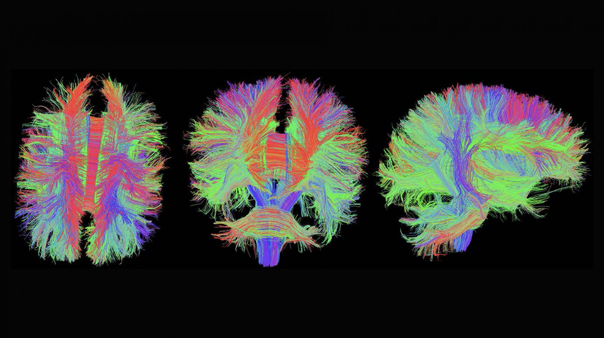
The objective of the GlyCORE Imaging Core is to promote and enhance the growth of glycoscience projects at the University of Mississippi, and throughout the mid-south region. The Core brings together new and existing advanced microscopes into a University-wide central platform, offering a wide range of advanced imaging techniques including laser scanning confocal microscopy and related beam parking experimental approaches, bright field and phase contrast microscopy, and computer-aided image analysis. The investigators will have access to and training in the use of advanced microscopy instrumentation, and assistance in the analysis of the resulting images. These images will provide new data and understandings on the roles of diverse carbohydrates in living systems.
As many advanced microscopic techniques are new to the University of Mississippi campus, the Imaging Core will develop a training program to inform and train new users on these new techniques. These techniques include, but are not limited to, the live cell techniques of FRET, FLIP and FRAP. Moreover, the GlyCORE Imaging Core will hold training sessions on how to properly get the most out of image analysis with instruction on the Leica Application Suite software, Image J/Fiji, the FARSIGHT toolkit for multi-dimensional microscopy, and Adobe Photoshop.
Imaging is essential for modern biological questions, and the GlyCORE Imaging Research Core will ensure glycoscience researchers at the University of Mississippi will have access to these tools. Through the availability of these instruments and training sessions, GlyCORE investigators will have a partner in advanced imaging techniques.
Prior to issuing a press release concerning any research outcome, please notify the PI of the award, Dr. Joshua Sharp, who will contact the Program Officer and NIGMS for the necessary coordination.
Imaging Core
The Biology Imaging Core is a shared and collaborative facility with the GlyCORE Imaging Core. The facility is designed to offer a full range of biological imaging capabilities. The imaging experts provide scientific advice and technical consultation for imaging experiment design and analysis.
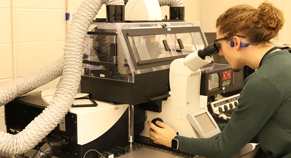
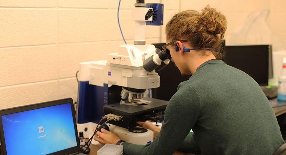
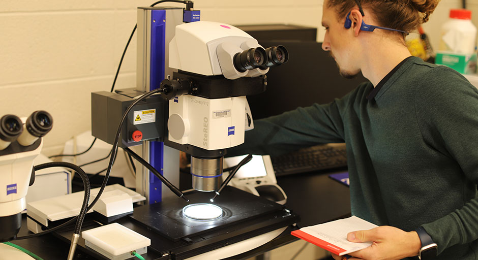
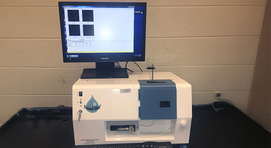
The core is equipped with a Leica SP8 inverted confocal microscope system with a temperature-controlled sample chamber, a Zeiss Axio M1 upright fluorescence microscope, and a Zeiss SteREO Discovery V12 modular stereo microscope, a Flowcam cytometer, and an image analysis workstation.
The facility provides users access to advanced microscopy instrumentation, training in the microscope operation for self-service and assistance in image analysis. Contact: Dr. John Adams, Shoemaker Hall Rm 114
Molecular Core
The Biology Molecular Biology Core is a self-service shared user facility that provides access to advanced instrumentation for the analysis of metabolites, proteins and nucleic acids.
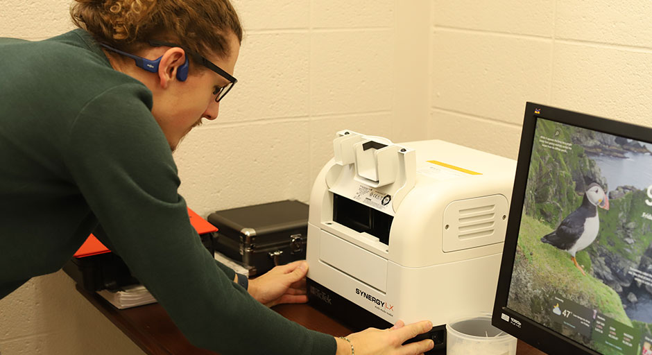
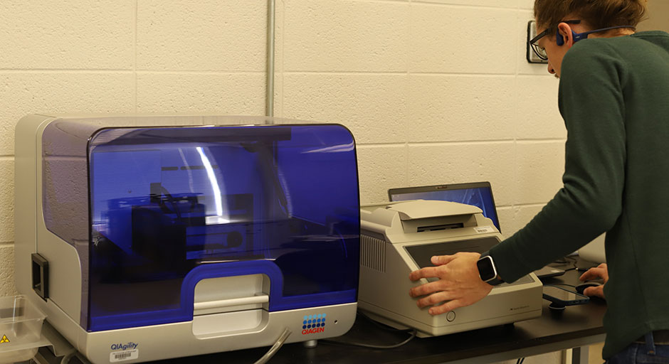
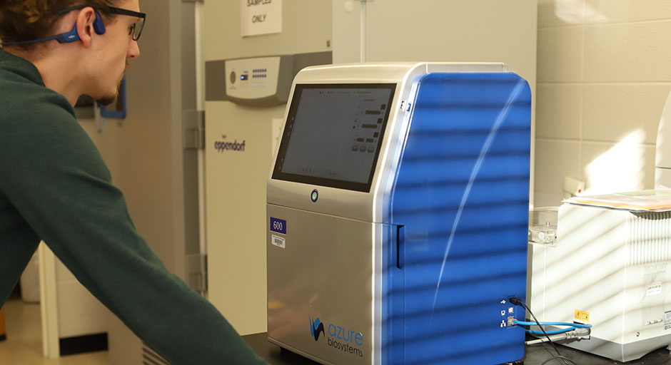
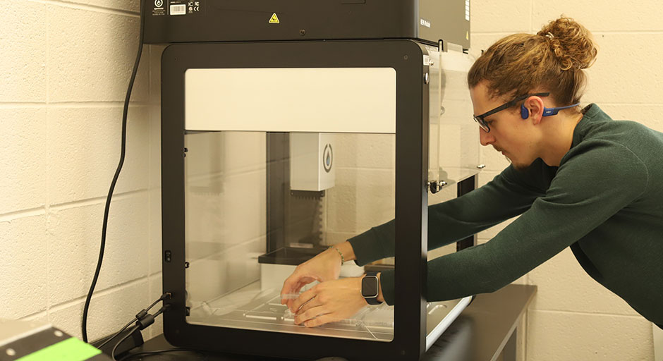
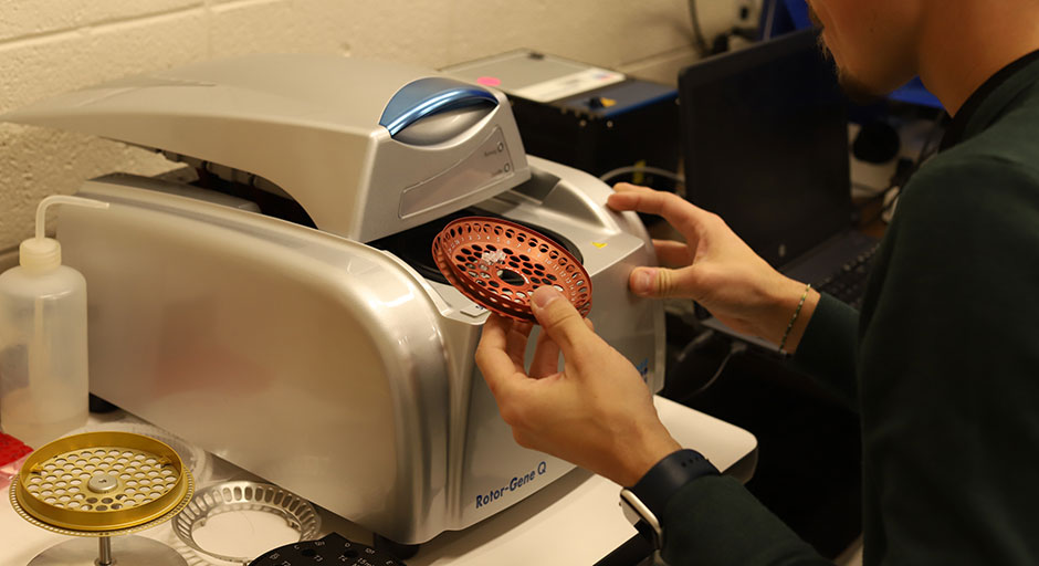
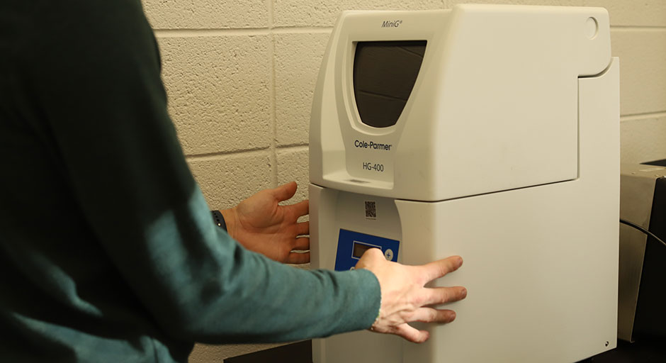
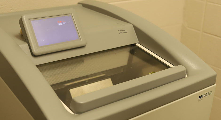
The core is equipped with an Azure C600 Advanced Imaging System for gel documentation and western blot, a Qiagen Rotor-Gene Q 5Plex real- time cycler, a QIAgility robotic system, a Biotek Synergy LX Multi-Mode Plate Reader, an OT-2 liquid handling robotic system, a Cryostat sample preparation station, and a SPEX GenoGrinder HG-400 MiniG.
Proper training is required before using any of the core instruments. Please contact the Biology Office to schedule training on any of the instruments.

